
Jaw Cysts: Diagnosis, Surgical Treatment, and What Pathology Tells You
Jaw cysts are fluid-filled cavities in the upper or lower jaw that develop from various tissue origins — most commonly from infected or non-vital teeth, sometimes from developmental remnants. Most are benign and treatable. The diagnostic workup matters because the type of cyst determines the surgical approach and the prognosis.
What jaw cysts are, and the main types
A cyst is a fluid-filled cavity lined by epithelium. In the jaws, the cyst lining is usually derived from epithelial remnants of tooth development. The main types relevant to UK adult patients:
Radicular (periapical) cyst
The most common — about 50–60% of jaw cysts. Develops at the tip of a tooth root with a non-vital (dead) pulp, almost always associated with previous decay or trauma. Pathology evolves through periapical granuloma → cyst over months to years.
Dentigerous cyst
Around 20% of cases. Forms around an impacted tooth (most often a wisdom tooth or upper canine) that has not erupted. The cyst surrounds the crown of the unerupted tooth.
Odontogenic keratocyst (OKC)
A more aggressive cyst with high recurrence rate. Now classified by WHO as keratocystic odontogenic tumour. Treatment requires more aggressive surgical approach and long-term follow-up.
Residual cyst
Persists in the bone after extraction of the tooth that originally caused the cyst.
Lateral periodontal cyst
Less common. Develops on the side of a vital tooth root, usually in lower premolars.
How they are detected
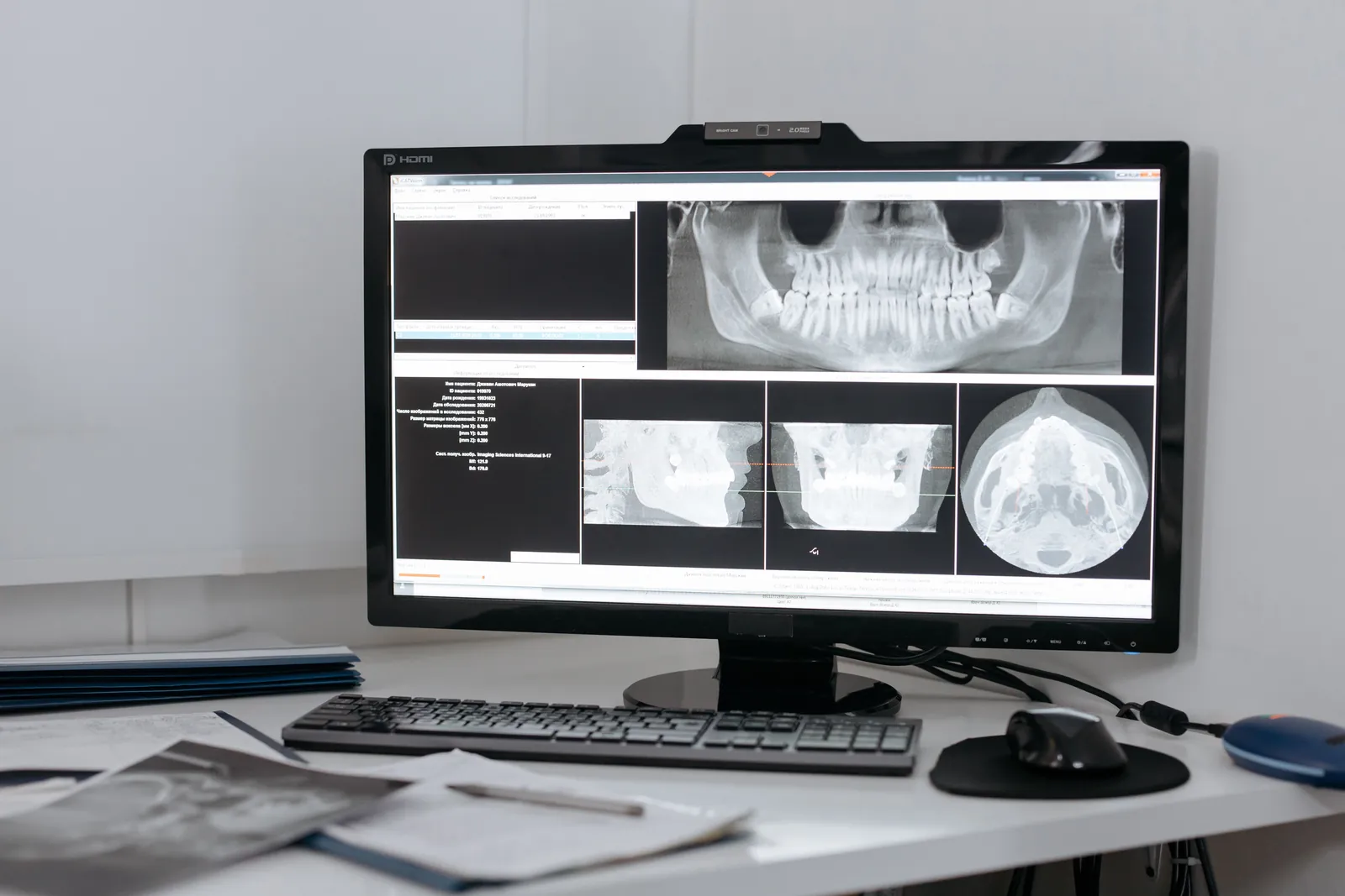
Most jaw cysts are silent — discovered as incidental findings on routine dental X-rays. The classic presentation: a circular or oval radiolucent area on a panoramic X-ray, well-defined borders, sometimes associated with tooth position changes.
When they are symptomatic, the signs include:
- Localised swelling that may have grown slowly over months.
- Tooth displacement or mobility.
- Numbness if the cyst is compressing a nerve.
- Sometimes pain or signs of acute infection if secondary infection has developed.
The diagnostic workup
Imaging:
- Panoramic X-ray (OPG): overview, often the first detection.
- Periapical X-ray: detail of relationship to tooth roots.
- CBCT: 3D assessment of cyst extent, relationship to nerves and sinuses, expansion of cortical bone. Essential for surgical planning of larger cysts.
Definitive diagnosis requires histological examination of the tissue removed at surgery — the X-ray gives strong likelihood, but pathology confirms.
Treatment approaches
Enucleation
Complete removal of the cyst lining via a small incision and bone window. Suitable for most small to medium cysts. Healing is straightforward; the bone defect fills with new bone over 6–12 months. Sutures removed at 7–10 days.
Marsupialisation
For large cysts, the cyst is opened and its lining sutured to the oral mucosa, creating a pouch that drains continuously. Over months, the cyst shrinks as new bone forms around the periphery. Final enucleation is performed when the cavity has reduced to manageable size. Used to avoid extensive bone removal in single-stage surgery.
Apicectomy with cystectomy
For radicular cysts associated with a tooth that can be saved, the apex of the tooth root is sealed (apicectomy) and the cyst removed. Our apicoectomy guide covers this combined procedure.
Extraction with cystectomy
For radicular cysts on non-restorable teeth, both the tooth and cyst are removed in one procedure. Implant replacement is planned for after bone healing.
OKC: the special case
Odontogenic keratocyst behaves more aggressively than other cysts:
- Higher recurrence rate (10–30% vs 1–5% for other cysts).
- Tends to grow along the bone rather than expand outward.
- Requires more aggressive treatment — enucleation plus peripheral ostectomy (drilling the bone surface beyond the cyst margin) or chemical fixation with Carnoy’s solution.
- Multiple OKCs can indicate Gorlin syndrome — a genetic condition with broader systemic implications.
- Long-term follow-up with annual X-rays for at least 5 years.
Recovery
- Day 0–3: moderate swelling and discomfort. Cold compress, OTC analgesia, soft food.
- Day 4–10: swelling reduces, sutures removed at 7–10 days.
- Week 2–6: gum tissue heals; bone fill begins.
- Month 3–12: progressive bone fill of the cavity. Routine X-ray follow-up at 6 and 12 months for routine cysts; more frequent for OKC.
For UK patients with detected jaw cyst
Most jaw cysts are detected on UK NHS X-rays during routine examination. NHS pathway: GP/general dental referral to oral surgery, NHS specialist assessment, surgical removal in hospital outpatient setting. Waiting times vary by region.
For UK patients considering Istanbul treatment for diagnosed cysts: send the existing imaging (X-rays, CBCT if available) for review before any travel. Some cysts are entirely manageable in routine NHS care; others may benefit from coordinated planning if combined with other treatments. Honest assessment of which scenario fits your case takes 24–48 hours after imaging review.
Frequently asked questions
Are jaw cysts dangerous?
Most are benign and treatable. Untreated cysts can grow over months to years, displacing teeth, expanding bone, and occasionally affecting nerves. Rare complications include malignant transformation (very low rate) or development into more aggressive lesions. Treatment is therefore appropriate even when symptoms are absent.
Will I need surgery for a cyst found on X-ray?
Usually yes, but the timing depends on the cyst type and size. Small radicular cysts may resolve with root canal treatment of the associated non-vital tooth. Larger cysts and other types typically need surgical removal. The diagnostic workup (CBCT, sometimes histology of an aspirated sample) determines the approach.
How long is recovery from cyst surgery?
For routine enucleation: 7–10 days for soft tissue healing, 6–12 months for full bone fill. Most patients return to normal activity within a week. Larger cysts treated by marsupialisation have a longer overall timeline (6+ months for shrinkage before final removal) but the impact on daily life is less.
Can a cyst come back after surgery?
Recurrence rate for routine cysts (radicular, dentigerous) is 1–5%. Odontogenic keratocyst (OKC) has higher recurrence — 10–30% — and requires more aggressive surgery and long-term follow-up with annual X-rays. The histology report from the original surgery determines the follow-up schedule.