
Dental X-Rays: Radiation Doses, Safety, and What Each Type Shows
Dental X-rays use far lower radiation doses than most patients assume — and the dose has dropped significantly since digital imaging replaced film. Understanding what each type of dental imaging shows and what the actual exposure is removes the worry from a routine diagnostic tool. Here is the realistic picture for UK patients.
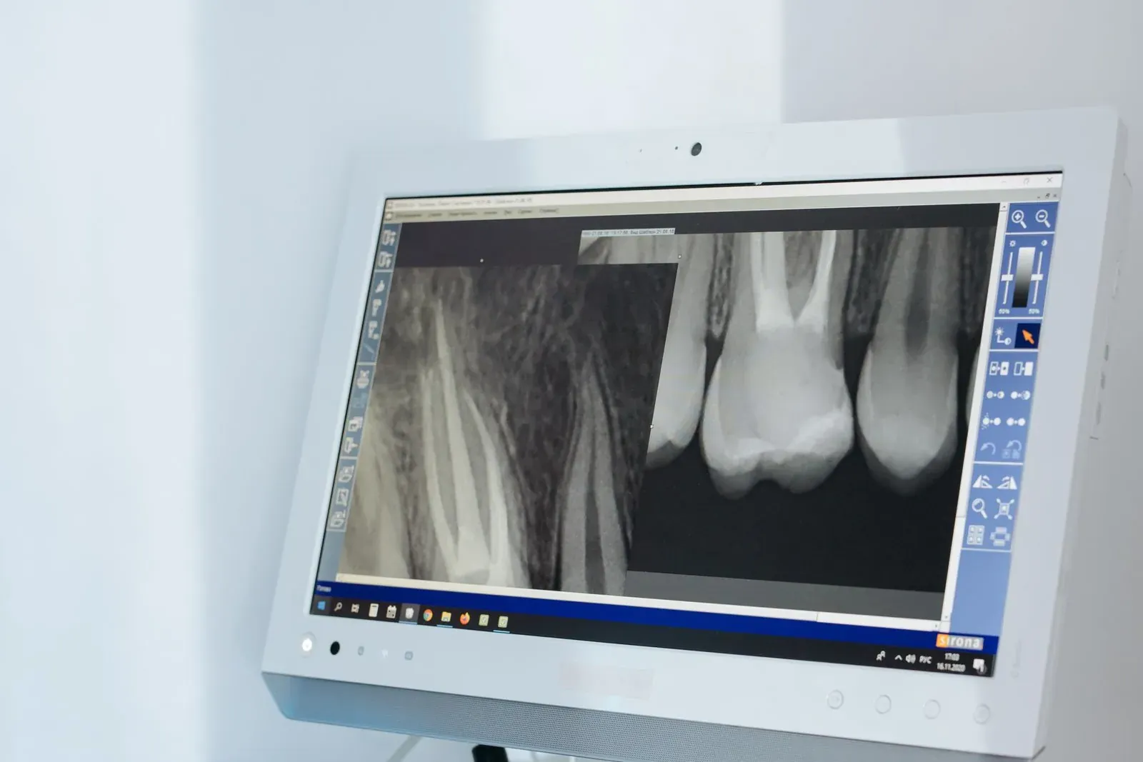
Putting dental X-ray doses in context
The radiation dose from a typical dental X-ray is measured in microsieverts (µSv). The relevant benchmarks:
- Single digital periapical X-ray: ~5 µSv.
- Two-bitewing set (routine 6-month or annual check): ~10 µSv.
- Full-mouth survey (14–18 X-rays for new patient assessment): ~30–40 µSv.
- Panoramic X-ray (OPG): ~15–20 µSv.
- CBCT 3D scan (small field of view, single tooth): ~50–100 µSv.
- CBCT 3D scan (full jaw, implant planning): ~200–400 µSv.
For comparison:
- Background radiation in the UK (everyone, every day): ~2,400 µSv/year, or about 6.6 µSv per day.
- One transatlantic flight (UK to New York return): ~80 µSv.
- Annual average chest X-ray: ~100 µSv.
- Smoker’s annual additional exposure (1 pack/day): ~25,000 µSv from polonium-210.
- CT abdomen/pelvis: ~10,000 µSv.
A single periapical X-ray equals about 18 hours of normal background exposure. A bitewing pair equals one transatlantic flight’s worth of cosmic radiation.
What each X-ray type actually shows
Bitewing
The standard cavity-detection X-ray. Shows the contact areas between teeth and the bone level around them. Most useful for detecting decay between teeth (where the toothbrush cannot see) and early periodontal bone loss. Routine annual or biannual check.
Periapical (PA)
Shows a single tooth from crown to root tip. Used for diagnosing pain in a specific tooth, evaluating root canal treatment, looking for periapical infection, planning extraction. Taken at the time of a clinical concern, not routinely.
Panoramic (OPG)
A single image showing all upper and lower teeth, both jaws, the temporomandibular joints, and the maxillary sinuses. Useful for new-patient screening, wisdom tooth evaluation, jaw fracture assessment. Less detail than periapical X-rays for individual teeth.
CBCT (3D cone beam)
A 3D volumetric scan. Shows bone in three dimensions, root anatomy in detail, position of the inferior dental nerve, sinus floor, and pathology that 2D X-rays cannot show. Used for implant planning, root canal anatomy that conventional X-rays cannot reveal, complex extraction planning, suspected fractures, and TMJ assessment. Higher dose than 2D X-rays but still a fraction of medical CT scans.
How modern dental X-rays are taken safely
- Digital sensors: 80% lower dose than the film systems they replaced.
- Rectangular collimation: beam narrowed to the size of the sensor (rather than a wider circular beam) further reduces stray radiation.
- Lead apron and thyroid collar: standard for routine X-rays. The thyroid collar specifically protects the thyroid gland from scatter radiation. Both are considered best practice in modern UK dentistry.
- Justification principle: X-rays are taken when there is a clinical reason, not on a fixed schedule. The Faculty of General Dental Practice (UK) selection criteria guide when X-rays are indicated.
- ALARA principle: “as low as reasonably achievable” — settings adjusted to the minimum dose that gives a diagnostic image.
Special situations
Pregnancy
Modern dental X-rays in pregnancy are safe with abdominal lead shielding when clinically necessary. Routine surveillance X-rays are usually deferred to after delivery; X-rays for active diagnostic concerns (toothache, abscess) are taken with appropriate shielding. The fetal exposure with lead shielding is essentially zero.
Children
Children receive proportionally smaller doses (settings adjusted for size). The cumulative lifetime exposure matters for pediatric patients, so X-rays are taken only when there is specific clinical indication. NHS guidance is conservative.
Cancer treatment history
Patients who have had radiotherapy to the head and neck region need careful planning of dental imaging. The treating oncologist’s input is appropriate for cumulative dose tracking.
The CBCT discussion specifically
CBCT is increasingly used in implant dentistry, complex root canal cases, and some orthodontic planning. The dose is higher than a periapical or panoramic but still substantially below medical CT. Key points:
- Used only when 2D imaging is insufficient for the clinical decision.
- Field of view (FOV) is restricted to the area of clinical interest, not the whole head.
- Modern CBCT machines have low-dose protocols specifically for dental applications.
- UK guidance from the Royal College of Radiologists supports CBCT for implant planning, complex endodontic cases, and other specific indications.
For UK patients planning implant treatment in Istanbul, the CBCT taken in the UK before travel is acceptable provided it is recent (within 12 months) and shows the relevant area at adequate resolution. Repeat scanning is avoided when not needed.
What the radiation actually does and does not cause
Ionising radiation can damage DNA, and at high doses this contributes to cancer risk. The doses involved in dental X-rays are far below the threshold where this becomes a measurable risk in epidemiological studies. Decades of population-level data show no detectable cancer increase from routine dental X-ray use at modern doses with appropriate shielding. The risk-benefit calculation for diagnostic dental X-rays is heavily favourable — the diagnosis they enable prevents far greater harm than the radiation involved.
Frequently asked questions
How much radiation is in a dental X-ray?
A single digital dental X-ray is approximately 5 µSv — roughly 18 hours of normal background radiation. A panoramic X-ray is 15–20 µSv. A CBCT scan is 50–400 µSv depending on field of view. For comparison, a transatlantic flight delivers around 80 µSv from cosmic radiation.
Are dental X-rays safe during pregnancy?
Yes when clinically necessary, with abdominal lead shielding. Routine X-rays are typically deferred to after delivery; X-rays for active dental problems (toothache, abscess) are taken when needed because the alternative — untreated infection — is a greater risk. Fetal exposure with shielding is essentially zero.
How often should I have dental X-rays?
There is no fixed schedule. Bitewings every 1–2 years for adults at moderate caries risk is typical; longer intervals for low-risk patients; shorter intervals for high-risk. Periapical X-rays are taken only when there is a specific clinical concern. The decision is the dentist’s based on individual risk, not a calendar rule.
Should I refuse dental X-rays to be safe?
Refusing X-rays without clinical justification means the dentist is working blind on issues that cannot be seen visually — early decay between teeth, periapical infections, bone loss patterns. The radiation risk is far smaller than the diagnostic risk of missing serious problems. If specific X-rays seem unnecessary, ask the dentist to explain the clinical reasoning rather than blanket-refusing.